Заключение
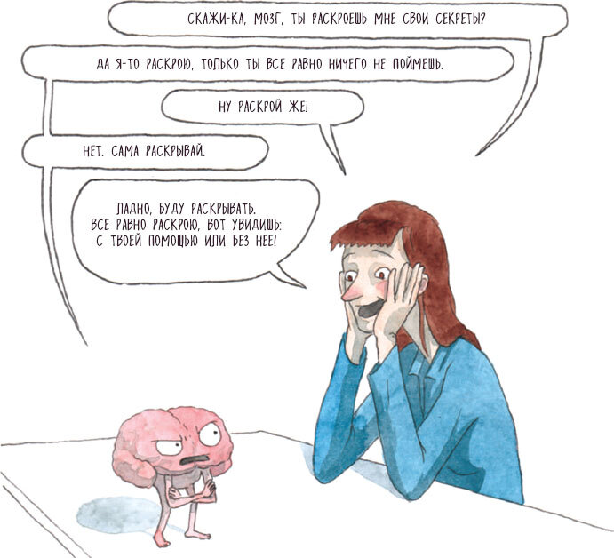
В последние годы исследования мозга переживают настоящий бум – идет ли речь о вопросах метафизического характера, касающихся сознания и индетерминизма, или о понимании и изучении некоторых механизмов работы мозга, чтобы сделать нас счастливее или энергичнее, или о борьбе с нейродегенеративными заболеваниями типа болезни Альцгеймера. Как бы то ни было, нам еще многое предстоит узнать. Получится ли это когда-нибудь? Вот что думает математик Иэн Стюарт (Ian Stewart): «Если бы наш мозг был устроен так просто, чтобы мы могли его понять, мы были бы устроены слишком просто, чтобы понять его» («If our brains were simple enough for us to understand them, we’d be so simple that we couldn’t»).

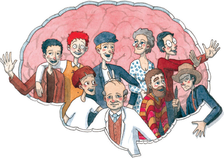
Ну а пока в этой книге мы поделились основными знаниями о мозге, которые есть на сегодняшний день, и воздали честь самым ярким личностям, внесшим весомый вклад в нейронауки. Пациенты, врачи или ученые – мы очень многим обязаны этим необыкновенным мозгам.

Библиография

СЮЗАННА. БЕССТРАШНЫЙ МОЗГ
Adolphs R. etal. (1994), «Impaired recognition of emotion in facial expressions following bilateral damage to the human amygdala», Nature, vol. 372, № 6507, p. 669–672.
Adolphs R. (2013), «The biology of fear», Current Biology, vol. 23, № 2, p. R79–R93.
Buchanan T. W. (2007), «Retrieval of emotional memories», Psychological bulletin, vol. 133, № 5, p. 761–779.
Damasio A. R. (1999), Le Sentiment même de soi. Corps, émotions, conscience, Paris, Odile Jacob.
Damasio A. R. (2006), L’Erreur de Descartes, Paris, Odile Jacob.
Feinstein J. S. et al. (2011), «The human amygdala and the induction and experience of fear», Current Biology, vol. 21, № 1, p. 34–38.
LeDoux J. (2003), «The emotional brain, fear and the amygdala», Cellular and Molecular Neurobiology, vol. 23, № 4–5, p. 727–738.
ГЕНРИ. ПАМЯТЬ ЗОЛОТОЙ РЫБКИ
Corkin S. (1984), «Lasting consequences of bilateral medial temporal lobectomy: Clinical course and experimental findings in H.M.», Seminars in Neurology, vol. 4, № 2, p. 249–259.
Corkin S. (2002), «What’s new with the amnesic patient H.M.?», Nature Reviews Neuroscience, vol. 3, № 2, p. 153–160.
Croisile B. (2009), Tout sur la mémoire, Paris, Odile Jacob.
Dossani R. H. et al. (2015), «The legacy of Henry Molaison (1926–2008) and the impact of his bilateral mesial temporal lobe surgery on the study of human memory», World Neurosurgery, vol. 84, № 4, p. 1127–1135.
Frankland P. W. & Bontempi B. (2005), «The organization of recent and remote memories», Nature Reviews Neuroscience, vol. 6, № 2, p. 119–130.
«Maladie d’Alzheimer», https://www.inserm.fr/information-en-sante/dossiers-information/ alzheimer-maladie.
«Mémoire», https://www.inserm.fr/information-en-sante/dossiers-information/memoire. Scoville W. B. & Milner B. (1957), «Loss of recent memory after bilateral hippocampal lesions», Journal of Neurology, Neurosurgery and Psychiatry, vol. 20, № 1, p. 11–21.
Squire L. R. (2009), «The legacy of patient H.M. for neuroscience», Neuron, vol. 61, № 1, p. 6–9.
ПЕНФИЛД. ОТКРЫТИЕ ГОМУНКУЛУСА
Bisi M. C. & Stagni R. (2016), «Development of gait motor control: What happens after asudden increase in height during adolescence?», BioMedical Engineering OnLine, vol. 15, p. 47.
Blum A. (2011), «Conversation au chevet de Wilder Penfield», CMAJ: Canadian Medical Association Journal, vol. 183, № 8, p. E477–E479.
Feindel W. et al. (2009), «Epilepsy surgery: Historical highlights 1909–2009», Epilepsia, vol. 50, p. 131–151.
Jeannerod M. (2006), «Plasticité du cortex moteur et récupération motrice», Motricité cérébrale. Réadaptation, neurologie du développement, vol. 27, № 2, p. 50–56.
Penfield W. (1954), «Mechanisms of voluntary movement», Brain, vol. 77, № 1, p. 1–17.
Penfield W. (1977), No Man Alone: A Surgeons Life, Little, Brown.
Penfield W. & Boldrey E. (1937), «Somatic motor and sensory representation in the cerebral cortex of man as studied by electrical stimulation», Brain, vol. 60, № 4, p. 389–443.
Roux F.-E. et al. (2003), «Cortical areas involved in virtual movement of phantom limbs: Comparison with normal subjects», Neurosurgery, vol. 53, № 6, p. 1342–1353.
Schlaug G. (2015), «Musicians and music making as a model for the study of brain plasticity», Progress in Brain Research, vol. 217, p. 37–55.
ДАВИД. ТРЕТИЙ МОЗГОВОЙ ГЛАЗ
Brindley G. S. etal. (1969), «Cortical blindness and the functions of the non-geniculate fibres of the optic tracts», Journal of Neurology, Neurosurgery & Psychiatry, vol. 32, № 4, p. 259–264.
Chokron S. (2013), «La cécité corticale: sémiologie, étiologie et perspectives de prise en charge neuropsychologique», Revue de neuropsychologie, vol. 5, № 1, p. 38–44.
«Le cerveau à tous les niveaux», http://lecerveau.mcgill.ca/.
Martinaud O. (2012), «Prosopagnosie et autres agnosies visuelles», Revue de neuropsychologie, vol. 4, № 4, p. 277–286.
Pöppel E. et al. (1973), «Residual visual function after brain wounds involving the central visual pathways in man», Nature, vol. 243, № 5405, p. 295–296.
Tamietto M. et al. (2009), «Unseen facial and bodily expressions trigger fast emotional reactions», Proceedings of the National Academy of Sciences, vol. 106, № 42, p. 17661–17666.
Weiskrantz L. et al. (1974), «Visual capacity in the hemianopic field following a restricted occipital ablation», Brain, vol. 97, № 4, p. 709–728.
ГОСПОДИН ТАН. ГОВОРЯЩИЙ МОЗГ
Bauchot R. (2009), «L’aphasie de Broca. Découverte par Paul Broca de la zone cérébrale du langage articulé», Bibnum. Textes fondateurs de la science.
Broca P. (1861), «Remarques sur le siège de la faculté du langage articulé, suivies d’une observation d’aphémie (perte de la parole)», Bulletin et mémoires de la Société anatomique de Paris, № 6, p. 330–357.
Broca P. (1868), Exposé des titres et travaux scientifiques de M. Paul Broca, candidat à l’Académie des sciences, section de médecine et de chirurgie, Paris, Hennuyer et fils.
Cohen L. (2003), L’Homme thermomètre. Le cerveau en pièces détachées, Paris, Odile Jacob. Domanski C. W. (2013), «Mysterious „Monsieur Leborgne“: The mystery of the famous patient in the history of neuropsychology is explained», Journal of the History of the Neurosciences, vol. 22, № 1, p. 47–52.
Dronkers N. F. et al. (2007), «Paul Broca’s historic cases: High resolution MR imaging of the brains of Leborgne and Lelong», Brain, vol. 130, № 5, p. 1432–1441.
Emmorey K. (2006), «The role of Broca’s area in sign language», in Y. Grodzinsky & K. Amunts, Broca’s Region, Oxford Scholar Online.
Planton S. & Démonet J-F. (2012), «Neurophysiologie du langage: apports de la neuro-imagerie et état des connaissances», Revue de neuropsychologie, vol. 4, № 4, p. 255–266.
Société d’anthropologie de Paris (1861), Bulletins de la Société d’anthropologie de Paris, vol. 2, p. 235–238.
Société d’anthropologie de Paris (1863), Bulletins de la Société d’anthropologie de Paris, vol. 4, p. 200–204.
Taglialatela J. P. et al. (2008), «Communicative signaling activates „Broca’s“ homolog in chimpanzees», Current biology, vol. 18, № 5, p. 343–348.
Thiebaut de Schotten M. et al. (2015), «From Phineas Gage and Monsieur Leborgne to H.M.: Revisiting disconnection syndromes», Cerebral Cortex, vol. 25, № 12, p. 4812–4827.
ФИНЕАС. АСОЦИАЛЬНЫЙ МОЗГ
Barker F. G. (1995), «Phineas among the phrenologists: the American crowbar case and nineteenth-century theories of cerebral localization», Journal of Neurosurgery, vol. 82, № 4, p. 672–682.
Besnard J. (2009), Contributions à l’étude des phénomènes de dépendance à l’environnement chez les patients cérébrolésés frontaux, thèse de doctorat en psychologie, université d’Angers. Derouesné C. (1979), «Frontal syndrome. I. Neuropsychological symptomatølogy», La Revue du praticien, vol. 29, № 35, p. 2809–2821.
Derouesné C. & Bakchine S. (2000), «Syndrome frontal», Encyclopédie médico-chirurgicale, № 17-035-B-10.
García-Molina A. (2012), «Phineas Gage and the enigma of the prefrontal cortex», Neurologia, vol. 27, № 6, p. 370–375.
Jeannerod M. (1992), «Organisation et désorganisation des fonctions mentales: le syndrome frontal», Revue de métaphysique et de morale, vol. 97, № 2, p. 235–253.
Le Doux J. (2003), Neurobiologie de la personnalité, Paris, Odile Jacob.
«Syndromes hémisphériques», https://www.cen-neurologie.fr/premier-cycle/semiologie-topographique/syndromes-peripheriques/syndromes-hemispheriques.
ДЖОЗЕФ. КОНФЛИКТ ДВУХ ПОЛУШАРИЙ
Corballis M. C. (2014), «Left brain, right brain: facts and fantasies», PLoS Biology, vol. 12, № 1.
Duboc V. et al. (2015), «Asymmetry of the brain: Development and implications», Annual Review of Genetics, vol. 49, № 1, p. 647–672.
Gazzaniga M. S. et al. (1962), «Some functional effects of sectioning the cerrebral commissures in man», Proceedings of the National Academy of Sciences of the United States of America, vol. 48, № 10, p. 1765–1769.
Gazzaniga M. S. et al. (1996), «Collaboration between the hemispheres of a callosotomy patient. Emerging right hemisphere speech and the left hemisphere interpreter», Brain, vol. 119, p. 1255–1262.
Gazzaniga M. S. (2000), «Cerebral specialization and interhemispheric communication. Does the corpus callosum enable the human condition?», Brain, vol. 123, № 7, p. 1293–1326.
Gazzaniga M. S. (2013), Le Libre arbitre et la science du cerveau, Paris, Odile Jacob. Gazzaniga M. S. & Smylie C. S. (1984), «Dissociation of language and cognition. A psychological profile of two disconnected right hemispheres», Brain, vol. 107, p. 145–153.
Gazzaniga M. S. & Sperry R. W. (1967), «Language after section of the cerebral commissures», Brain, vol. 90, № 1, p. 131–148.
Nielsen J. A. et al. (2013), «An evaluation of the left-brain vs right-brain hypothesis with resting state functional connectivity magnetic resonance imaging», PloS One, vol. 8, № 8, p. e71275.
Sidtis J. J. et al. (1981), «Cognitive interaction after staged callosal section: Evidence for transfer of semantic activation», Science, vol. 212, № 4492, p. 344–346.
Vallortigara G. et al. (1999), «Possible evolutionary origins of cognitive brain lateralization», Brain Research Reviews, vol. 30, № 2, p. 164–175.
Volz L. J. & Gazzaniga M. S. (2017), «Interaction in isolation: 50 years of insights from split-brain research», Brain, vol. 140, № 7, p. 2051–2060.
Wolman D. (2012), «The split brain: A tale of two halves», Nature News, vol. 483, № 7389, p. 260.
«Severed corpus callosum», https://www.youtube.com/watch?v=lfGwsAdS9Dc.
ПОЛЬ. СИНДРОМ НЕГЛЕКТА
Brain W. R. (1941), «A form of visual disorientation resulting from lesions of the right cerebral hemisphere», Proceedings of the Royal Society of Medicine, vol. 34, № 12, p. 771–776.
Chabert A.-L. (2014), Transformer le «handicap» ou l’invention d’un usage détourné du monde. Essai de cheminement conceptuel à partir d’expériences de vie, thèse de doctorat en philosophie des sciences, université Paris-Diderot.
Jewell G. & McCourt M. E. (2000), «Pseudoneglect: A review and meta-analysis of performance factors in line bisection tasks», Neuropsychologia, vol. 38, № 1, p. 93–110.
Marshall J. C. & Halligan P. W. (1988), «Blindsight and insight in visuo-spatial neglect», Nature, vol. 336, № 6201, p. 766.
Ochando A. & Zago L. (2018), «What are the contributions of handedness, sighting dominance, hand used to bisect, and visuospatial line processing to the behavioral line bisection bias?», Frontiers in Psychology, vol. 9, p. 1688.
Parton A. et al. (2004), «Hemispatial neglect», Journal of Neurology, Neurosurgery and Psychiatry, vol. 75, № 1, p. 13–21.
Paterson A. & Zangwill O. L. (1944), «Disorders of visual space perception associated with lesions of the right cerebral hemisphere», Brain, vol. 67, № 4, p. 331–358.
Paterson A. & Zangwill O. L. (1945), «A case of topographical disorientation associated with a unilateral cerebral lesion», Brain, vol. 68, № 3, p. 188–212.
Rossetti Y. et al. (1998), «Prism adaptation to a rightward optical deviation rehabilitates left hemispatial neglect», Nature, vol. 395, № 6698, p. 166–169.
Sacks O. (2014), L’Homme qui prenait sa femme pour un chapeau. Et autres récits cliniques, Paris, Seuil, «Points».
Seron X. & Van der Linden M. (2014), Traité de neuropsychologie clinique de l’adulte, tome 1: Évaluation, De Boeck Supérieur.
СОЛОМОН. ЕДИНСТВО ОЩУЩЕНИЙ
Ameisen J.-C., «Le mnémoniste», Sur les épaules de Darwin, France Inter, 27 mai 2017, 54 min.
Luria A. (1992), Une prodigieuse mémoire, Paris, Delachaux et Niestlé.
Hubbard E. M. & Ramachandran V. S. (2005), «Neurocognitive mechanisms of synesthesia», Neuron, vol. 48, № 3, p. 509–520.
Rothen N. et al. (2012), «Enhanced memory ability: Insights from synaesthesia», Neuroscience & Biobehavioral Reviews, vol. 36, № 8, p. 1952–1963.
Rouw R. et al. (2011), «Brain areas involved in synaesthesia: A review», Journal of Neuropsychology, vol. 5, № 2, p. 214–242.
Shams L. & Seitz A. R. (2008), «Benefits of multisensory learning», Trends in Cognitive Sciences, vol. 12, № 11, p. 411–417.
КОЕ-ЧТО ЕЩЕ
Agid Y. & Magistretti P. (2018), L’Homme glial. Une révolution dans les sciences du cerveau, Paris, Odile Jacob.
Feuillet L. et al. (2007), «Brain of a white-collar worker», The Lancet, vol. 370, № 9583, p. 262.
Herculano-Houzel S. (2009), «The human brain in numbers: A linearly scaled-up primate brain», Frontiers in Human Neuroscience, vol. 3.
Jeannerod M. (2007), L’Homme sans visage et autres récits de neurologie quotidienne, Paris, Odile Jacob.
Naccache L. & Naccache K. (2018), Parlez-vous cerveau?, Paris, Odile Jacob.
Ramachandran V. S. & Blakeslee S. (2002), Le Fantôme intérieur, Paris, Odile Jacob.
«Are there really as many neurons in the human bain as stars in the milky way?», https:// www.nature.com/scitable/blog/brain-metrics/are_there_really_as_many/.

